

Is The Act Of Passively Receiving And Registering The Sounds That Reach Your Ears.
| Ear | |
|---|---|
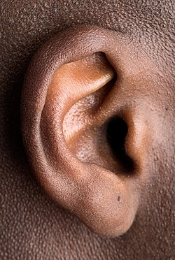 The outer portion of the homo ear | |
| Details | |
| System | Auditory system |
| Identifiers | |
| Latin | Auris |
| MeSH | D004423 |
| NeuroLex ID | birnlex_1062 |
| TA98 | A01.1.00.005 A15.3.00.001 |
| TA2 | 6861 |
| FMA | 52780 |
| Anatomical terminology [edit on Wikidata] | |
How sounds make their way from the source to the homo brain
The ear is the organ that enables hearing and, in mammals, body rest using the vestibular system. In mammals, the ear is usually described every bit having three parts—the outer ear, the middle ear and the inner ear. The outer ear consists of the pinna and the ear canal. Since the outer ear is the but visible portion of the ear in most animals, the word "ear" often refers to the external part alone.[1] The eye ear includes the tympanic cavity and the three ossicles. The inner ear sits in the bony labyrinth, and contains structures which are fundamental to several senses: the semicircular canals, which enable residuum and eye tracking when moving; the utricle and saccule, which enable residuum when stationary; and the cochlea, which enables hearing. The ears of vertebrates are placed somewhat symmetrically on either side of the head, an arrangement that aids sound localisation.
The ear develops from the kickoff pharyngeal pouch and six small-scale swellings that develop in the early embryo called otic placodes, which are derived from ectoderm.
The ear may exist affected by disease, including infection and traumatic impairment. Diseases of the ear may lead to hearing loss, tinnitus and balance disorders such as vertigo, although many of these conditions may also be affected past damage to the encephalon or neural pathways leading from the ear.
The ear has been adorned past earrings and other jewelry in numerous cultures for thousands of years, and has been subjected to surgical and cosmetic alterations.
Structure
The human ear consists of 3 parts—the outer ear, eye ear and inner ear.[two] The ear culvert of the outer ear is separated from the air-filled tympanic cavity of the middle ear past the eardrum. The middle ear contains the three small basic—the ossicles—involved in the manual of audio, and is continued to the pharynx at the nasopharynx, via the pharyngeal opening of the Eustachian tube. The inner ear contains the otolith organs—the utricle and saccule—and the semicircular canals belonging to the vestibular system, as well equally the cochlea of the auditory system.[2]
Outer ear
The outer ear is the external portion of the ear and includes the fleshy visible pinna (besides called the auricle), the ear canal, and the outer layer of the eardrum (as well called the tympanic membrane).[2] [3]
The pinna consists of the curving outer rim chosen the helix, the inner curved rim called the antihelix, and opens into the ear canal. The tragus protrudes and partially obscures the ear culvert, as does the facing antitragus. The hollow region in front end of the ear canal is called the concha. The ear canal stretches for about 1inch (two.5cm). The first part of the canal is surrounded by cartilage, while the second function well-nigh the eardrum is surrounded by os. This bony part is known as the auditory bulla and is formed by the tympanic office of the temporal bone. The skin surrounding the ear canal contains ceruminous and sebaceous glands that produce protective ear wax. The ear culvert ends at the external surface of the eardrum.[3]
Two sets of muscles are associated with the outer ear: the intrinsic and extrinsic muscles. In some mammals, these muscles can adjust the direction of the pinna.[3] In humans, these muscles take piddling or no effect.[iv] The ear muscles are supplied by the facial nerve, which also supplies awareness to the skin of the ear itself, likewise every bit to the external ear crenel. The great auricular nerve, auricular nerve, auriculotemporal nerve, and lesser and greater occipital nerves of the cervical plexus all supply sensation to parts of the outer ear and the surrounding peel.[3]
The pinna consists of a single piece of elastic cartilage with a complicated relief on its inner surface and a fairly smooth configuration on its posterior surface. A tubercle, known every bit Darwin'south tubercle, is sometimes present, lying in the descending part of the helix and respective to the ear-tip of mammals. The earlobe consists of areola and adipose tissue.[5] The symmetrical arrangement of the 2 ears allows for the localisation of sound. The brain accomplishes this by comparing arrival-times and intensities from each ear, in circuits located in the superior olivary complex and the trapezoid bodies which are connected via pathways to both ears.[half-dozen]
Middle ear

The middle ear lies between the outer ear and the inner ear. It consists of an air-filled cavity called the tympanic cavity and includes the 3 ossicles and their attaching ligaments; the auditory tube; and the round and oval windows. The ossicles are three modest bones that part together to receive, amplify, and transmit the sound from the eardrum to the inner ear. The ossicles are the malleus (hammer), incus (anvil), and the stapes (stirrup). The stapes is the smallest named os in the body. The middle ear also connects to the upper throat at the nasopharynx via the pharyngeal opening of the Eustachian tube.[3] [7]
The three ossicles transmit sound from the outer ear to the inner ear. The malleus receives vibrations from sound pressure on the eardrum, where it is continued at its longest part (the manubrium or handle) by a ligament. Information technology transmits vibrations to the incus, which in plow transmits the vibrations to the small stapes os. The broad base of the stapes rests on the oval window. Equally the stapes vibrates, vibrations are transmitted through the oval window, causing motility of fluid within the cochlea.[3]
The round window allows for the fluid within the inner ear to move. As the stapes pushes the secondary tympanic membrane, fluid in the inner ear moves and pushes the membrane of the round window out by a corresponding amount into the heart ear. The ossicles help amplify sound waves past about 15–twenty times.[2]
Inner ear

The inner ear sits within the temporal os in a complex crenel called the bony labyrinth. A central area known as the foyer contains two small-scale fluid-filled recesses, the utricle and saccule. These connect to the semicircular canals and the cochlea. There are three semicircular canals angled at right angles to each other which are responsible for dynamic balance. The cochlea is a spiral shell-shaped organ responsible for the sense of hearing. These structures together create the membranous labyrinth.[8]
The bony labyrinth refers to the bony compartment which contains the membranous labyrinth, contained within the temporal os. The inner ear structurally begins at the oval window, which receives vibrations from the incus of the eye ear. Vibrations are transmitted into the inner ear into a fluid chosen endolymph, which fills the membranous labyrinth. The endolymph is situated in two vestibules, the utricle and saccule, and eventually transmits to the cochlea, a spiral-shaped structure. The cochlea consists of three fluid-filled spaces: the vestibular duct, the cochlear duct, and the tympanic duct.[3] Hair cells responsible for transduction—changing mechanical changes into electric stimuli are present in the organ of Corti in the cochlea.[eight]
Blood supply
The blood supply of the ear differs according to each role of the ear.
The outer ear is supplied by a number of arteries. The posterior auricular artery provides the majority of the blood supply. The anterior auricular arteries provide some supply to the outer rim of the ear and scalp backside information technology. The posterior auricular artery is a straight branch of the external carotid artery, and the anterior auricular arteries are branches from the superficial temporal avenue. The occipital avenue too plays a role.[8]
The middle ear is supplied by the mastoid branch of either the occipital or posterior auricular arteries and the deep auricular artery, a branch of the maxillary avenue. Other arteries which are present but play a smaller role include branches of the middle meningeal artery, ascending pharyngeal artery, internal carotid artery, and the artery of the pterygoid canal.[8]
The inner ear is supplied by the anterior tympanic branch of the maxillary artery; the stylomastoid branch of the posterior auricular artery; the petrosal branch of middle meningeal artery; and the labyrinthine artery, arising from either the anterior inferior cerebellar avenue or the basilar artery.[8]
Function
Hearing
Sound waves travel through the outer ear, are modulated past the middle ear, and are transmitted to the vestibulocochlear nerve in the inner ear. This nerve transmits information to the temporal lobe of the brain, where it is registered as sound.
Sound that travels through the outer ear impacts on the eardrum, and causes it to vibrate. The iii ossicles bones transmit this audio to a second window (the oval window) which protects the fluid-filled inner ear. In detail, the pinna of the outer ear helps to focus a sound, which impacts on the eardrum. The malleus rests on the membrane, and receives the vibration. This vibration is transmitted along the incus and stapes to the oval window. Two pocket-sized muscles, the tensor tympani and stapedius, likewise help modulate noise. The two muscles reflexively contract to dampen excessive vibrations. Vibration of the oval window causes vibration of the endolymph within the vestibule and the cochlea.[9]
The inner ear houses the appliance necessary to change the vibrations transmitted from the exterior world via the middle ear into signals passed along the vestibulocochlear nerve to the brain. The hollow channels of the inner ear are filled with liquid, and contain a sensory epithelium that is studded with hair cells. The microscopic "hairs" of these cells are structural poly peptide filaments that project out into the fluid. The hair cells are mechanoreceptors that release a chemic neurotransmitter when stimulated. Sound waves moving through fluid flows against the receptor cells of the organ of Corti. The fluid pushes the filaments of individual cells; move of the filaments causes receptor cells to become open to receive the potassium-rich endolymph. This causes the cell to depolarise, and creates an activity potential that is transmitted along the spiral ganglion, which sends information through the auditory portion of the vestibulocochlear nerve to the temporal lobe of the encephalon.[9]
The human ear tin by and large hear sounds with frequencies between 20 Hz and 20 kHz (the audio range). Sounds outside this range are considered infrasound (below 20 Hz)[10] or ultrasound (above twenty kHz)[11] Although hearing requires an intact and performance auditory portion of the central nervous organisation too as a working ear, human deafness (farthermost insensitivity to sound) most commonly occurs because of abnormalities of the inner ear, rather than in the nerves or tracts of the central auditory system.
Balance
Providing balance, when moving or stationary, is also a central function of the ear. The ear facilitates two types of balance: static balance, which allows a person to feel the effects of gravity, and dynamic rest, which allows a person to sense dispatch.
Static residue is provided past two ventricles, the utricle and the saccule. Cells lining the walls of these ventricles comprise fine filaments, and the cells are covered with a fine gelled layer. Each cell has fifty–lxx modest filaments, and one large filament, the kinocilium. Within the gelatinous layer lie otoliths, tiny formations of calcium carbonate. When a person moves, these otoliths shift position. This shift alters the positions of the filaments, which opens ion channels within the cell membranes, creating depolarisation and an action potential that is transmitted to the brain along the vestibulocochlear nerve.[nine] [12]
Dynamic balance is provided through the 3 semicircular canals. These three canals are orthogonal (at right angles) to each other. At the cease of each culvert is a slight enlargement, known as the ampulla, which contains numerous cells with filaments in a central area called the cupula. The fluid in these canals rotates according to the momentum of the caput. When a person changes acceleration, the inertia of the fluid changes. This affects the pressure on the cupula, and results in the opening of ion channels. This causes depolarisation, which is passed as a betoken to the encephalon forth the vestibulocochlear nervus.[nine] Dynamic remainder also helps maintain eye tracking when moving, via the vestibulo–ocular reflex.
Development
During embryogenesis the ear develops as 3 distinct structures: the inner ear, the centre ear and the outer ear.[13] Each structure originates from a different germ layer: the ectoderm, endoderm and mesenchyme.[14] [xv]
Inner ear

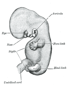
The otic placode visible on this sketch of a developing embryo.
After implantation, effectually the second to 3rd calendar week the developing embryo consists of three layers: endoderm, mesoderm and ectoderm. The first function of the ear to develop is the inner ear,[fifteen] which begins to class from the ectoderm effectually the 22nd day of the embryo'southward development.[xiv] Specifically, the inner ear derives from ii thickenings called otic placodes on either side of the caput. Each otic placode recedes beneath the ectoderm, forms an otic pit and so an otic vesicle.[16] This unabridged mass will eventually become surrounded by mesenchyme to form the bony labyrinth.[16] [17]
Around the 33rd mean solar day of development, the vesicles brainstorm to differentiate. Closer to the back of the embryo, they form what volition get the utricle and semicircular canals. Closer to the forepart of the embryo, the vesicles differentiate into a rudimentary saccule, which will somewhen go the saccule and cochlea. Part of the saccule volition eventually give ascension and connect to the cochlear duct. This duct appears approximately during the 6th week and connects to the saccule through the ductus reuniens.[14]
Every bit the cochlear duct'due south mesenchyme begins to differentiate, 3 cavities are formed: the scala vestibuli, the scala tympani and the scala media.[xiv] [17] Both the scala vestibuli and the scala tympani comprise an extracellular fluid called perilymph. The scala media contains endolymph.[17] A set of membranes called the vestibular membrane and the basilar membrane develop to separate the cochlear duct from the vestibular duct and the tympanic duct, respectively.[14]
Parts of the otic vesicle in turn class the vestibulocochlear nerve.[xviii] These form bipolar neurons which supply sensation to parts of the inner ear (namely the sensory parts of the semicircular canals, macular of the utricle and saccule, and organ of Corti). The nerve begins to form effectually the 28th day.[xvi]
- Molecular regulation
Most of the genes responsible for the regulation of inner ear germination and its morphogenesis are members of the homeobox gene family such as Pax, Msx and Otx homeobox genes. The development of inner ear structures such equally the cochlea is regulated by Dlx5/Dlx6, Otx1/Otx2 and Pax2, which in turn are controlled past the principal gene Shh. Shh is secreted by the notochord.[19]
Centre ear
The eye ear and its components develop from the first and second pharyngeal arches.[16] The tympanic cavity and auditory tube develop from the showtime part of the pharyngeal pouch between the first two arches in an area which will also go along to develop the throat. This develops as a structure called the tubotympanic recess.[16] The ossicles (malleus, incus and stapes) normally appear during the first half of fetal development. The first ii (malleus and incus) derive from the showtime pharyngeal arch and the stapes derives from the 2nd.[14] All three ossicles develop from the neural crest.[16] Somewhen cells from the tissue surrounding the ossicles volition experience apoptosis and a new layer of endodermal epithelial will constitute the germination of the tympanic cavity wall.[xiv] [15]
Outer ear
The ear develops in the lower cervix region and moves up as the mandible develops.
Unlike structures of the inner and middle ear, which develop from pharyngeal pouches, the ear culvert originates from the dorsal portion of the first pharyngeal cleft.[14] [16] It is fully expanded past the end of the 18th week of development.[17] The eardrum is made upwardly of three layers (ectoderm, endoderm and connective tissue). The pinna originates as a fusion of half-dozen hillocks. The first three hillocks are derived from the lower part of the first pharyngeal arch and form the tragus, crus of the helix, and helix, respectively. The final three hillocks are derived from the upper part of the second pharyngeal arch and course the antihelix, antitragus, and earlobe.[fourteen] [sixteen] [17] The outer ears develop in the lower neck. As the mandible forms they move towards their concluding position level with the optics.[13] [18]
Clinical significance
Hearing loss

Perforation

Fluid in the heart ear cavity
Complications of otitis media that can lead to hearing loss, as seen on otoscope.
Hearing loss may exist either partial or total. This may exist a result of injury or damage, built disease, or physiological causes. When hearing loss is a result of injury or damage to the outer ear or middle ear, information technology is known as conductive hearing loss. When deafness is a effect of injury or harm to the inner ear, vestibulochoclear nerve, or brain, information technology is known as sensorineural hearing loss.
Causes of conductive hearing loss include an ear culvert blocked by ear wax, ossicles that are stock-still together or absent, or holes in the eardrum. Conductive hearing loss may besides result from middle ear inflammation causing fluid build-up in the ordinarily air-filled space, such as by otitis media. Tympanoplasty is the general proper noun of the operation to repair the center ear's eardrum and ossicles. Grafts from muscle fascia are normally used to rebuild an intact eardrum. Sometimes artificial ear basic are placed to substitute for damaged ones, or a disrupted ossicular chain is rebuilt in order to carry sound effectively.
Hearing aids or cochlear implants may be used if the hearing loss is astringent or prolonged. Hearing aids work by amplifying the sound of the local environs and are best suited to conductive hearing loss.[20] Cochlear implants transmit the sound that is heard as if information technology were a nervous signal, bypassing the cochlea. Active middle ear implants send audio vibrations to the ossicles in the middle ear, bypassing whatsoever not-operation parts of the outer and middle ear.
Congenital abnormalities
Anomalies and malformations of the pinna are common. These anomalies include chromosome syndromes such as ring 18. Children may besides present cases of abnormal ear canals and low ear implantation.[15] In rare cases no pinna is formed (atresia), or is extremely pocket-sized (microtia). Small pinnae can develop when the auricular hillocks practise non develop properly. The ear canal can fail to develop if it does not channelise properly or if there is an obstruction.[15] Reconstructive surgery to treat hearing loss is considered every bit an option for children older than five,[21] with a cosmetic surgical process to reduce the size or modify the shape of the ear is called an otoplasty. The initial medical intervention is aimed at assessing the baby'due south hearing and the condition of the ear culvert, likewise equally the centre and inner ear. Depending on the results of tests, reconstruction of the outer ear is done in stages, with planning for whatever possible repairs of the rest of the ear.[22] [23] [24]
Approximately one out of one chiliad children suffer some type of congenital deafness related to the development of the inner ear.[25] Inner ear congenital anomalies are related to sensorineural hearing loss and are generally diagnosed with a computed tomography (CT) browse or a magnetic resonance imaging (MRI) browse.[21] Hearing loss problems likewise derive from inner ear anomalies because its evolution is separate from that of the middle and external ear.[xv] Centre ear anomalies can occur because of errors during head and neck development. The commencement pharyngeal pouch syndrome assembly heart ear anomalies to the malleus and incus structures every bit well equally to the non-differentiation of the annular stapedial ligament. Temporal bone and ear canal anomalies are also related to this structure of the ear and are known to be associated with sensorineural hearing loss and conductive hearing loss.[21]
Vertigo
Vertigo refers to the inappropriate perception of motion. This is due to dysfunction of the vestibular system. One common type of vertigo is benign paroxysmal positional vertigo, when an otolith is displaced from the ventricles to the semicircular canal. The displaced otolith rests on the cupola, causing a sensation of move when there is none. Ménière's disease, labyrinthitis, strokes, and other infective and congenital diseases may too effect in the perception of vertigo.[26]
Injury
- Outer ear
Injuries to the external ear occur fairly frequently, and tin can leave minor to major deformity. Injuries include: laceration, avulsion injuries, burn and repeated twisting or pulling of an ear, for bailiwick or torture.[27] Chronic damage to the ears tin cause cauliflower ear, a common condition in boxers and wrestlers in which the cartilage around the ears becomes lumpy and distorted owing to persistence of a haematoma around the perichondrium, which can impair claret supply and healing.[28] Owing to its exposed position, the external ear is susceptible to frostbite[29] as well equally skin cancers, including squamous-cell carcinoma and basal-cell carcinomas.[30]
- Centre ear
The ear drum may go perforated in the event of a large sound or explosion, when diving or flying (chosen barotrauma), or by objects inserted into the ear. Another common cause of injury is due to an infection such as otitis media.[31] These may cause a discharge from the ear called otorrhea,[32] and are often investigated by otoscopy and audiometry. Treatment may include watchful waiting, antibiotics and maybe surgery, if the injury is prolonged or the position of the ossicles is affected.[33] Skull fractures that become through the part of the skull containing the ear structures (the temporal bone) can also cause damage to the center ear.[34] A cholesteatoma is a cyst of squamous peel cells that may develop from birth or secondary to other causes such as chronic ear infections. It may impair hearing or crusade dizziness or vertigo, and is normally investigated by otoscopy and may require a CT scan. The handling for cholesteatoma is surgery.[35]
- Inner ear
There are ii principal damage mechanisms to the inner ear in industrialised gild, and both injure pilus cells. The first is exposure to elevated sound levels (noise trauma), and the second is exposure to drugs and other substances (ototoxicity). A big number of people are exposed to sound levels on a daily basis that are likely to lead to pregnant hearing loss.[36] The National Institute for Occupational Safety and Health has recently published research on the estimated numbers of persons with hearing difficulty (11%) and the per centum of those that tin can be attributed to occupational noise exposure (24%).[37] Furthermore, according to the National Health and Nutrition Examination Survey (NHANES), approximately xx-two million (17%) U.s.a. workers reported exposure to hazardous workplace noise.[38] Workers exposed to hazardous noise farther exacerbate the potential for developing racket-induced hearing loss when they do not wear hearing protection.
Tinnitus
Tinnitus is the hearing of audio when no external sound is nowadays.[39] While oftentimes described as a ringing, it may as well sound like a clicking, hiss or roaring.[40] Rarely, unclear voices or music are heard.[41] The sound may be soft or loud, low pitched or high pitched and appear to be coming from 1 ear or both.[xl] Most of the time, information technology comes on gradually.[41] In some people, the audio causes depression, anxiety, or concentration difficulties.[xl]
Tinnitus is not a disease but a symptom that can effect from a number of underlying causes. One of the most common causes is noise-induced hearing loss. Other causes include: ear infections, disease of the middle or claret vessels, Ménière's disease, brain tumors, emotional stress, exposure to sure medications, a previous head injury, and earwax.[40] [42] Information technology is more mutual in those with depression and feet.[41]
Lodge and culture

Stretching of the earlobe and diverse cartilage piercings
The ears have been ornamented with jewelry for thousands of years, traditionally by piercing of the earlobe. In ancient and modern cultures, ornaments have been placed to stretch and enlarge the earlobes, assuasive for larger plugs to be slid into a large fleshy gap in the lobe. Vehement of the earlobe from the weight of heavy earrings, or from traumatic pull of an earring (for example, past snagging on a sweater), is adequately common.[43]
Injury to the ears has been present since Roman times as a method of reprimand or punishment – "In Roman times, when a dispute arose that could not be settled amicably, the injured party cited the name of the person thought to exist responsible before the Praetor; if the offender did not appear within the specified time limit, the complainant summoned witnesses to make statements. If they refused, as often happened, the injured party was allowed to elevate them by the ear and to pinch them hard if they resisted. Hence the French expression "se faire tirer l'oreille", of which the literal meaning is "to have i's ear pulled" and the figurative pregnant "to take a lot of persuading". We use the expression "to tweak (or pull) someone's ears" to hateful "inflict a punishment"."[27]
The pinnae have an outcome on facial appearance. In Western societies, protruding ears (present in most five% of ethnic Europeans) have been considered unattractive, particularly if asymmetric.[44] The first surgery to reduce the projection of prominent ears was published in the medical literature past Ernst Dieffenbach in 1845, and the start case report in 1881.[45]

Pointy ears are a feature of some creatures in folklore such equally the French croquemitaine, Brazilian curupira[46] or Japanese earth spider.[47] It has been a feature of characters on art equally quondam every bit that of Ancient Greece[48] and medieval Europe.[49] Pointy ears are a common characteristic of many creatures in the fantasy genre,[50] including elves,[51] [52] [53] faeries,[54] [55] pixies,[56] hobbits,[57] or orcs.[58] They are a feature of creatures in the horror genre, such as vampires.[59] [60] Pointy ears are also found in the scientific discipline fiction genre; for example among the Vulcan and Romulan races of the Star Trek universe[61] and the Nightcrawler graphic symbol from the X-Men universe.[62]
Georg von Békésy was a Hungarian biophysicist born in Budapest, Republic of hungary. In 1961, he was awarded the Nobel Prize in Physiology or Medicine for his inquiry on the function of the cochlea in the mammalian hearing organ.[63]
The Vacanti mouse was a laboratory mouse that had what looked like a human ear grown on its back. The "ear" was actually an ear-shaped cartilage structure grown by seeding cow cartilage cells into a biodegradable ear-shaped mold and so implanted nether the peel of the mouse; and then the cartilage naturally grew by itself.[64] It was adult equally an alternative to ear repair or grafting procedures and the results met with much publicity and controversy in 1997.[65] [66]
Other animals


The pinna helps directly sound through the ear canal to the eardrum. The circuitous geometry of ridges on the inner surface of some mammalian ears helps to sharply focus sounds produced past prey, using echolocation signals. These ridges can exist regarded equally the acoustic equivalent of a fresnel lens, and may be seen in a broad range of animals, including the bat, aye-aye, lesser galago, bat-eared fox, mouse lemur and others.[67] [68] [69]
Some large primates such every bit gorillas and orang-utans (and likewise humans) have undeveloped ear muscles that are non-functional vestigial structures, still are nevertheless large enough to be hands identified.[70] An ear muscle that cannot motion the ear, for any reason, has lost that biological function. This serves as show of homology between related species. In humans, there is variability in these muscles, such that some people are able to move their ears in various directions, and information technology has been said that it may exist possible for others to gain such movement by repeated trials.[lxx] In such primates, the inability to motility the ear is compensated for mainly past the power to easily plough the head on a horizontal airplane, an ability which is not mutual to almost monkeys—a function once provided past one structure is now replaced by another.[71]
In some animals with mobile pinnae (like the horse), each pinna can be aimed independently to meliorate receive the sound. For these animals, the pinnae help localise the direction of the sound source.

Analogy by
Charles Darwin, 1868
The ear, with its blood vessels shut to the surface, is an essential thermoregulator in some land mammals, including the elephant, the pull a fast one on, and the rabbit.[72] At that place are v types of ear wagon in domestic rabbits, some of which have been bred for exaggerated ear length[73]—a potential wellness risk that is controlled in some countries.[74] Abnormalities in the skull of a one-half-lop rabbit were studied by Charles Darwin in 1868. In marine mammals, Earless seals are one of three groups of Pinnipedia.
Invertebrates
Only vertebrate animals have ears, though many invertebrates notice sound using other kinds of sense organs. In insects, tympanal organs are used to hear distant sounds. They are located either on the caput or elsewhere, depending on the insect family.[75] The tympanal organs of some insects are extremely sensitive, offer acute hearing beyond that of virtually other animals. The female cricket fly Ormia ochracea has tympanal organs on each side of her belly. They are connected by a thin bridge of exoskeleton and they office similar a tiny pair of eardrums, merely, because they are linked, they provide acute directional information. The fly uses her "ears" to detect the telephone call of her host, a male cricket. Depending on where the vocal of the cricket is coming from, the fly's hearing organs will reflect at slightly unlike frequencies. This departure may be equally little as l billionths of a 2nd, just it is enough to allow the fly to home in directly on a singing male cricket and parasitise it.[76]
Simpler structures allow other arthropods to detect near-field sounds. Spiders and cockroaches, for example, have hairs on their legs which are used for detecting sound. Caterpillars may also have hairs on their torso that perceive vibrations[77] and allow them to respond to sound.
Run across also
- Hear, hear
- Hearing test
- Righting reflex
References
- ^ "Ear". Oxford Dictionary . Retrieved 25 February 2016.
- ^ a b c d Standring, Susan (2008). Borley, Neil R. (ed.). Greyness's Anatomy: The Anatomical Basis of Clinical Do (40 ed.). Edinburgh: Churchill Livingstone/Elsevier. pp. Chapter 36. "External and centre ear", 615–631. ISBN978-0-443-06684-9. Archived from the original on 10 March 2014.
- ^ a b c d east f g Drake, Richard L.; Vogl, Wayne; Tibbitts, Adam W.M. Mitchell; illustrations past Richard; Richardson, Paul (2005). Greyness's anatomy for students. Philadelphia: Elsevier/Churchill Livingstone. pp. 855–856. ISBN978-0-8089-2306-0.
- ^ Moore KL, Dalley AF, Agur AM (2013). Clinically Oriented Anatomy, 7th ed. Lippincott Williams & Wilkins. pp. 848–849. ISBN978-1-4511-8447-one.
- ^ Stenström, J. Sten: Deformities of the ear; In: Grabb, W., C., Smith, J.S. (Edited): "Plastic Surgery", Piddling, Brown and Visitor, Boston, 1979, ISBN 0-316-32269-5 (C), ISBN 0-316-32268-7 (P)
- ^ Purves, D. (2007). Neuroscience (fourth ed.). New York: Sinauer. pp. 332–336. ISBN978-0-87893-697-7.
- ^ Mitchell, Richard L. Drake, Wayne Vogl, Adam W.M. (2005). Gray's anatomy for students. Philadelphia: Elsevier. p. 858. ISBN978-0-8089-2306-0.
- ^ a b c d e Standring, Susan (2008). Borley, Neil R. (ed.). Grayness's Anatomy: The Anatomical Footing of Clinical Practice (40 ed.). Edinburgh: Churchill Livingstone/Elsevier. pp. Chapter 37. "Inner ear", 633–650. ISBN978-0-443-06684-9.
- ^ a b c d Hall, Arthur C. Guyton, John E. (2005). Textbook of medical physiology (11th ed.). Philadelphia: W.B. Saunders. pp. 651–657. ISBN978-0-7216-0240-0.
- ^ Greinwald, John H. Jr Doctor; Hartnick, Christopher J. MD The Evaluation of Children With Sensorineural Hearing Loss. Archives of Otolaryngology – Head & Neck Surgery. 128(one):84–87, Jan 2002
- ^ "Definition of "ultrasound" | Collins English language Lexicon". www.collinsdictionary.com . Retrieved 20 March 2016.
- ^ Hall, Arthur C. Guyton, John E. (2005). Textbook of medical physiology (11th ed.). Philadelphia: W.B. Saunders. pp. 692–694. ISBN978-0-7216-0240-0.
- ^ a b Moore, Keith L. (2009). Fundamentos de Anatomía con Orientación Clínica. pp. 1021–1035.
- ^ a b c d e f k h i Sadler, T.West. (2010). Embriología Médica. pp. 321–327.
- ^ a b c d e f Moore, Keith L. (2008). Embriología Clínica. pp. 477–482.
- ^ a b c d east f g h Standring, Susan (2008). Borley, Neil R. (ed.). Gray's Anatomy: The Anatomical Basis of Clinical Practice (40 ed.). Edinburgh: Churchill Livingstone/Elsevier. pp. Chapter 38. "Development of the ear", 651–653. ISBN978-0-443-06684-9.
- ^ a b c d due east UNSW Embryology. Hearing-Inner Ear Evolution. Archived from the original on 30 September 2012. Retrieved 20 April 2013.
- ^ a b Drake, Richard Fifty.; Wayne, A.; Mitchell, Adam (2010). GRAY Anatomía para estudiantes. pp. 854–871.
- ^ Chatterjee, Sumantra; Kraus, Petra; Luftkin, Thomas (2010). "A symphony of inner ear developmental control genes". BMC Genetics. eleven: 68. doi:10.1186/1471-2156-11-68. PMC2915946. PMID 20637105.
- ^ "Hearing Aids". National institute of deafness and other communication disorders . Retrieved twenty March 2016.
- ^ a b c Kliegman; Behrman; Jenson (2007). "367". Nelson Textbook of Pedriatics.
- ^ Lam SM. Edward Talbot Ely: male parent of artful otoplasty. [Biography. Historical Commodity. Journal Commodity] Archives of Facial Plastic Surgery. vi(one):64, 2004 Jan–February.
- ^ Siegert R. Combined reconstruction of built auricular atresia and severe microtia. [Evaluation Studies. Periodical Article] Laryngoscope. 113(xi):2021–2027; discussion 2028–2029, 2003 Nov.
- ^ Trigg DJ. Applebaum EL. Indications for the surgical repair of unilateral aural atresia in children. [Review] [33 refs] [Journal Commodity. Review], American Journal of Otology. 19(5):679–684; discussion 684–686, 1998 September
- ^ Lalwani, A.Chiliad. (2009). Diagnóstico y tratamiento en Otorrinolaringología. Cirugía de Cabeza y Cuello. pp. 624–752.
- ^ Britton, the editors Nicki R. Colledge, Brian R. Walker, Stuart H. Ralston; illustrated past Robert (2010). Davidson's principles and practice of medicine (21st ed.). Edinburgh: Churchill Livingstone/Elsevier. pp. 1151–1171. ISBN978-0-7020-3084-0.
- ^ a b Alexandru, Florin (30 January 2004). "Ear Injuries" (PDF). Council of Europe.
- ^ "Ear Injury – Injuries and Poisoning". Merck Manuals Consumer Version . Retrieved 25 February 2016.
- ^ Colledge, Nicki (2010). Davidson's Principles and Exercise of Medicine. Churchill Livingstone. p. 102.
- ^ "The Ears, A High Risk Area for Pare Cancer". www.skincancer.org. Archived from the original on iv March 2016. Retrieved 25 Feb 2016.
- ^ "Ruptured Eardrum: Symptoms, Treatments, and Recovery". WebMD . Retrieved 25 February 2016.
- ^ "How should I evaluate a draining ear?". Medscape . Retrieved 25 February 2016.
- ^ "Traumatic Perforation of the Tympanic Membrane – Ear, Olfactory organ, and Throat Disorders". Merck Manuals Professional person Edition . Retrieved 25 February 2016.
- ^ "Evaluation and management of center ear trauma". www.uptodate.com . Retrieved 25 February 2016.
- ^ "Cholesteatoma: MedlinePlus Medical Encyclopedia". www.nlm.nih.gov . Retrieved 25 February 2016.
- ^ Senate Public Works Committee, Noise disturbance and Abatement Act of 1972, Due south. Rep. No. 1160, 92nd Cong. 2nd session.
- ^ Tak SW, Calvert GM, "Hearing Difficulty Attributable to Employment by Industry and Occupation: An Analysis of the National Health Interview Survey – United States, 1997 to 2003," J. Occup. Env. Med. 2008, fifty:46–56
- ^ Tak, SW; Davis, RR; Calvert, GM (2009). "Exposure to Chancy Workplace Racket and Use of Hearing Protection Devices Amongst U.s. WOrkers, 1999–2004". Am. J. Ind. Med. 52 (5): 358–371. doi:10.1002/ajim.20690. PMID 19267354.
- ^ Levine, RA; Oron, Y (2015). Tinnitus. Handbook of Clinical Neurology. Vol. 129. pp. 409–431. doi:ten.1016/B978-0-444-62630-i.00023-viii. ISBN978-0-444-62630-1. PMID 25726282.
- ^ a b c d "Tinnitus". September 2014. Retrieved 22 May 2015.
- ^ a b c Baguley, D; McFerran, D; Hall, D (nine November 2013). "Tinnitus" (PDF). Lancet. 382 (9904): 1600–1607. doi:x.1016/S0140-6736(13)60142-7. PMID 23827090. Archived from the original (PDF) on xi April 2018. Retrieved thirty June 2019.
- ^ Han BI, Lee HW, Kim TY, Lim JS, Shin KS (March 2009). "Tinnitus: characteristics, causes, mechanisms, and treatments". J Clin Neurol. five (ane): 11–19. doi:10.3988/jcn.2009.5.one.11. PMC2686891. PMID 19513328.
About 75% of new cases are related to emotional stress as the trigger factor rather than to precipitants involving cochlear lesions.
{{cite journal}}: CS1 maint: uses authors parameter (link) - ^ Deborah Southward. Sarnoff; Robert H. Gotkin; Joan Swirsky (2002). Instant Beauty: Getting Gorgeous on Your Lunch Suspension. St. Martin's Press. ISBN0-312-28697-X.
- ^ Thomas, J. Regan (2010). Advanced Therapy in Facial Plastic and Reconstructive Surgery. PMPH-The states. p. 513. ISBN978-1-60795-011-0.
- ^ Miloro, Michael; Ghali, Thousand.Due east.; Larsen, Peter; Waite, Peter (2004). "Chapter 71. Otoplastic surgery for the protruding ear.". Peterson'due south Principles of Oral and Maxillofacial Surgery. PMPH-USA. ISBN978-1-55009-234-9.
- ^ Theresa Bane (2013). Encyclopedia of Fairies in World Folklore and Mythology. McFarland. p. 91. ISBN978-0-7864-7111-9.
- ^ Laurence Bush-league (2001). Asian Horror Encyclopedia: Asian Horror Culture in Literature, Manga, and Folklore. iUniverse. p. 43. ISBN978-1-4697-1503-2.
- ^ Johann Joachim Winckelmann (1850). The History of Ancient Fine art Among the Greeks. Chapman. p. 80.
- ^ Alixe Bovey (2002). Monsters and Grotesques in Medieval Manuscripts. University of Toronto Press. p. 38. ISBN978-0-8020-8512-2.
- ^ J. Peffer (2012). DragonArt Collector's Edition: Your Ultimate Guide to Drawing Fantasy Fine art. IMPACT. p. 28. ISBN978-ane-4403-2417-8.
- ^ Michael J. Tresca (2010). The Evolution of Fantasy Role-Playing Games. McFarland. p. 34. ISBN978-0-7864-6009-0.
- ^ David Okum (2006). Manga Fantasy Madness: Over 50 Basic Lessons for Drawing Warriors, Wizards, Monsters and more. IMPACT. p. 31. ISBN1-60061-381-0.
- ^ Sirona Knight (7 June 2005). The Complete Idiot'southward Guide to Elves and Fairies. DK Publishing. p. 171. ISBN978-i-4406-9638-one.
- ^ John Michael Greer (1 September 2011). Monsters. Llewellyn Worldwide. p. 107. ISBN978-0-7387-1600-8.
- ^ Christopher Hart (2008). Amazing Fantasy Worlds: The Ultimate Guide to Cartoon Take a chance Fantasy Art. Watson-Guptill Publications. p. 103. ISBN978-0-8230-1472-9.
- ^ John Hamilton (ane August 2011). Elves and Fairies. ABDO. p. 23. ISBN978-1-60453-215-nine.
- ^ Misha Kavka; Jenny Lawn; Mary Paul (2006). Gothic Nz: The Darker Side of Kiwi Culture. Otago Academy Press. p. 111. ISBN978-ane-877372-23-0.
- ^ Lisa Hopkins (1 January 2010). Screening the Gothic. University of Texas Press. p. 202. ISBN978-0-292-77959-iv.
- ^ Noah William Isenberg (xiii August 2013). Weimar Cinema: An Essential Guide to Archetype Films of the Era. Columbia University Press. pp. 96–. ISBN978-0-231-50385-three.
- ^ Ken Gelder (2000). The Horror Reader. Psychology Press. p. 27. ISBN978-0-415-21356-1.
- ^ Henry Jenkins III; Tara McPherson; Jane Shattuc (2 Jan 2003). Hop on Pop: The Politics and Pleasures of Popular Culture. Duke University Press. p. 119. ISBN0-8223-8350-0.
- ^ William Irwin; Rebecca Housel; J. Jeremy Wisnewski (18 May 2009). X-Men and Philosophy: Astonishing Insight and Uncanny Argument in the Mutant X-Poetry. John Wiley & Sons. p. 189. ISBN978-0-470-73036-ii.
- ^ Stevens, S.S. (September 1972). "Georg von Békésy". Physics Today. 25 (9): 78–eighty. Bibcode:1972PhT....25i..78S. doi:ten.1063/1.3071029. Archived from the original on 24 September 2013.
- ^ Cao, Y.; Vacanti, J.P.; Paige, One thousand.T.; Upton, J.; Vacanti, C.A. (1997). "Transplantation of chondrocytes utilizing a polymer-cell construct to produce tissue-engineered cartilage in the shape of a human ear". Plastic and Reconstructive Surgery. 100 (2): 297–302, discussion 303–304. doi:ten.1097/00006534-199708000-00001. PMID 9252594. S2CID 41167703.
- ^ Goodyear, Dana. "The Stress Test". New Yorker. Retrieved 23 March 2016.
- ^ Karin Sellberg, Lena Wånggren (2016). Corporeality and Culture: Bodies in Movement. Routledge. pp. 75–76. ISBN978-i-317-15924-7.
- ^ Pavey, C.R.; Burwell, C.J. (1998). "Bat Predation on Eared Moths: A Exam of the Allotonic Frequency Hypothesis". Oikos. 81 (1): 143–151. doi:10.2307/3546476. JSTOR 3546476.
- ^ "The Bat's Ear as a Diffraction Grating". Archived from the original on xviii April 2012. Retrieved 27 October 2011.
- ^ Kuc, R. (2009). "Model predicts bat pinna ridges focus loftier frequencies to course narrow sensitivity beams". The Journal of the Acoustical Club of America. 125 (v): 3454–3459. Bibcode:2009ASAJ..125.3454K. doi:10.1121/ane.3097500. PMID 19425684.
- ^ a b Darwin, Charles (1871). The Descent of Man, and Selection in Relation to Sexual activity. John Murray: London.
- ^ Mr. St. George Mivart, Elementary Beefcake, 1873, p. 396. Two ears provide stereo imaging that the brain can use to develop a 3-dimensional sound field.
- ^ Fayez, I.; Marai, 1000.; Alnaimy, A.; Habeeb, M. (1994). Baselga, M.; Marai, I.F.Thou. (eds.). "Thermoregulation in Rabbits" (PDF). Rabbit Production in Hot Climates. Cahiers Options Méditerranéennes. Zaragoza: CIHEAM – International Centre for Advanced Mediterranean Agronomic Studies. 8: 33–41.
- ^ "Longest ears on a rabbit". Guinness Globe Records . Retrieved ix February 2018.
- ^ Whitman, Bob D. (October 2004). Domestic Rabbits & Their Histories: Breeds of the World. Leawood KS: Leathers Publishing. ISBN978-1-58597-275-3.
- ^ Yack, JE; Fullard, JH (1993). "What is an insect ear?". Ann. Entomol. Soc. Am. 86 (6): 677–682. doi:ten.1093/aesa/86.six.677.
- ^ Piper, Ross (2007), Extraordinary Animals: An Encyclopedia of Curious and Unusual Animals, Greenwood Press.
- ^ Scoble, Thou.J. 1992. The Lepidoptera: Form, function, and multifariousness. Oxford University Press
External links

Source: https://en.wikipedia.org/wiki/Ear
Posted by: maginnisentlead.blogspot.com
0 Response to "Is The Act Of Passively Receiving And Registering The Sounds That Reach Your Ears."
Post a Comment